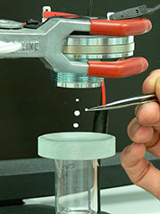
Ultrasonic tweezers for life-changing medical advances
Researchers from the Universities of Bristol and Southampton have helped to develop pioneering ‘tweezers’ that use ultrasound beams to grip and manipulate tiny clusters of cells, which could lead to life-changing medical advances, such as better cartilage implants that reduce the need for knee replacement operations.
The team worked with test equipment maker Agilent, Crystapol International, the Defence Science and Technology Laboratory (DSTL), Leica Genetix, Loadpoint, Piezo Composite Transducers (PCT), Weidlinger Associates and IKTS-Fraunhofer.
Using ultrasonic sound fields, cartilage cells taken from a patient’s knee can be levitated for weeks in a nutrient-rich fluid. This means the nutrients can reach every part of the culture’s surface and, combined with the stimulation provided by the ultrasound, enables the cells to grow and to form better implant tissue than when grown on a glass petri dish.
The tweezers, developed with £3.6m of Engineering and Physical Sciences Research Council (EPSRC) funding, involve multiple, tiny beams of ultrasonic waves that, in a typical device, point into a 10 mm-diameter chamber from all around. With the aid of a powerful microscope to monitor the procedure, the forces generated by the waves can then be manipulated so that they nudge cells into the required position, turn them around, or hold them firmly in place. The ‘four year Electronic Sonotweezers: Particle Manipulation with Ultrasonic Arrays’ programme started in 2009 and also includes researchers from the Universities of Dundee and Glasgow.
By holding the cells in the required position firmly but gently, the tweezers can also mould the growing tissue into exactly the right shape so that the implant is truly fit-for-purpose when inserted into the patient’s knee.
“Ultrasonic tweezers can provide what is, in effect, a zero-gravity environment perfect for optimising cell growth,” said Professor Martyn Hill, Head of the Engineering Sciences Unit at the University of Southampton, who led the cartilage tissue engineering work in collaboration with colleagues Dr Peter Glynne-Jones, New Frontiers Fellow in Engineering Sciences, Dr Rahul Tare, a Lecturer in Musculoskeletal Science and Bioengineering, and Professor Richard Oreffo, a Professor of Musculoskeletal Science. “As well as levitating cells, the tweezers can make sure that the cell agglomerates maintain a flat shape ideal for nutrient absorption. They can even gently massage the agglomerates in a way that encourages cartilage tissue formation.”
Professor Bruce Drinkwater of Bristol University, who co-ordinated the programme, said: “Ultrasonic tweezers have all kinds of possible uses in bioscience, nanotechnology and more widely across industry. They offer big advantages over optical tweezers relying on light waves and also over electromagnetic methods of cell manipulation; for example, they have a complete absence of moving parts and can manipulate not just one or two cells at a time but clusters up to 1mm across – a scale that makes them very suitable for applications like tissue engineering.”
The research programme has also shown that ultrasonic tweezers can be used to build up cell tissue layer by layer, which could for instance, help to reconstruct nerve tissue after severe trauma such as limb amputation.
This research will enable ultrasonic tweezer technology to be refined and miniaturised and specific uses to be explored and developed in the next few years. The first real-world applications, in sectors such as bioscience and electronics, could potentially be developed within around five years.
http://www.bristol.ac.uk/physics/research/nanophysics/facilities/tweezers)
Related articles
Bristol hospitals team with NPL to make breast cancer detection more reliable
Initial tests show promising results for new ultrasonic screening technique
The main hospitals in Bristol are working with the National Physical Laboratory on a initial trial of a new, potentially more reliable, technique for screening breast cancer using ultrasound. The team at NPL are now looking to develop the technique into a clinical device.
“Our initial results are very promising. Whilst it’s early days, we’re very excited about its potential and with the right funding, support and industry partners, we may well have something here which could have a huge and positive impact on cancer diagnosis and the lives of many thousands of women,” said Dr Bajram Zeqiri, who leads the project at NPL.
The project was funded by the research arm of the NHS, the National Institute of Health Research, under its Invention for Innovation funding stream and co-funded by the NPL Strategic Research Programme. University Hospitals Bristol NHS Foundation Trust is a leading UK centre in breast screening using ultrasound and partnered with NPL on the initial tests. They are now working on a demonstrator and will look to work with a manufacturer to commercialise the technology.
Around 46,000 women are diagnosed with breast cancer in the UK every year, mostly using breast screening based on X-ray mammography. Only about 30% of suspicious lesions turn out to be malignant. Each lesion must be confirmed by invasive biopsies, estimated to cost the NHS £35 million per year. Ionising radiation also has the potential to cause cancer, which limits the use of X-rays to single screenings of at risk groups, such as women over 50 through the National Breast Screening Programme.
There is a compelling need to develop improved, ideally non-ionising, methods of detecting breast lesions and solid masses. Improved diagnosis would reduce unnecessary biopsies and consequent patient trauma from being wrongly diagnosed.
Ultrasound ticks many of the boxes: it is safe, low cost, and already extensively used in trusted applications such as foetal scanning. However the quality of the images is not yet good enough for reliable diagnoses.
Part of the problem lies with the current detectors used. Different biological tissues have different sound speeds, and this affects the time taken for sound waves to arrive at the detector. This can distort the arriving waves, in extreme cases causing cancellation them to cancel each other out. This results in imaging errors, such as suggesting abnormal inclusions where there may be none.
The new method works by detecting the intensity of ultrasonic waves. Intensity is converted to heat that is then sensed by a thin membrane of pyroelectric film, which generates a voltage output dependant on the temperature rise. Imaging detectors based on this new principle should be much less susceptible to the effects caused by the uneven sound speed in tissues.
This technique, when used in a Computed Tomography (CT) configuration, should produce more accurate images of tissue properties and so provide better identification of breast tissue abnormalities. The aim of tomography is to produce a cross-section map of the tissue, which describes how the acoustic properties vary across the tissue. Using this map, it is possible to identify abnormal inclusions.
An initial feasibility project has proved the concept by testing single detectors using purpose-built artefacts. These artefacts were designed to include well-defined structures, enabling the new imaging method to be compared with more conventional techniques. The results confirmed that the new detectors generated more reliable maps of the internal structure of the artefacts than existing techniques.
NPL is now seeking funding to develop the work further. They hope to produce a demonstrator using a full array of 20 sensors, which should allow more rapid scanning and move the idea towards a system which might eventually be used clinically. It is hoped that this will provide both a suitable resolution and fast enough scanning to become a viable replacement for current clinical scanners.
Related articles
- Breast cancer screening benefits are oversold and harms could be greater than thought (dailymail.co.uk)
- What Is Breast Thermography? (everydayhealth.com)
- Breast Cancer Diagnosis (everydayhealth.com)
- Ultrasound for Breast Cancer Detection (everydayhealth.com)


